About fifty thousand people each year in the United States experience corneal hazing, the impairment of vision by scar tissue on the cornea. Also occurring in staggering numbers, traumatic brain injuries affect about two million people yearly worldwide. In order to decrease those numbers, Professor Orwin and her lab have been working on two major projects involving injured cells in the body. One project focuses on rehabilitating injured cornea cells, and the other looks at brain cells damaged from the inflammatory response triggered after brain damage.
For the cornea team, the techniques implemented include electromagnetic stimulation, mechanical stimulation, and the alteration of matrix stiffness. Electromagnetic stimulation involves exposing the damaged cells to light, which actually helps with their tissue repair. The team has tried a variety of wavelengths of light, and they have found that blue light is especially effective in electromagnetic stimulation of the injured cornea cells. For mechanical stimulation, they use a bioreactor to simulate a healthy environment for the cell in the body, thus aiding their rehabilitation. Changing the matrix stiffness also involves placing the cell in a healthy environment, but that environment is generated by an electrified needle. The needle ejects protein in a desired structure, and the arrangement of that structure affects how the injured cells will behave. The rehabilitation of the cornea has not been studied as often as other topics in biomedical research, so Prof. Orwin and her students are very excited about the research they have been performing in this area.
For the brain patch team, the cells of interest are brain cells called astrocytes which are important to the wound-healing process in the brain. However, they use rabbit corneal fibroblasts in the lab in place of astrocytes due to their greater robustness. Still, they use some astrocytes to create an accurate brain wound model. The astrocytes require special coatings to be applied and special densities for seeding, meaning they need to be tested only in specific concentrations. Nevertheless, the student team finds working with them intriguing, as senior Risa Egerter describes, “We culture them from a fresh sample, meaning a baby rat’s brain, which I think is kind of cool.”
As one of the largest labs on campus, Prof. Orwin’s lab has been performing biomedical research for thirteen years. Senior Travis Beckman, who is working on the cornea project, praises his research experience: “Research allows you to fall back on self-motivation. Your frustration gets channeled into productivity.” Prof. Orwin also thinks highly of the students in her lab, saying, “Research is a good way to get faculty and students working together closely. The students build a community amongst themselves that adds to their sense of teamwork.”
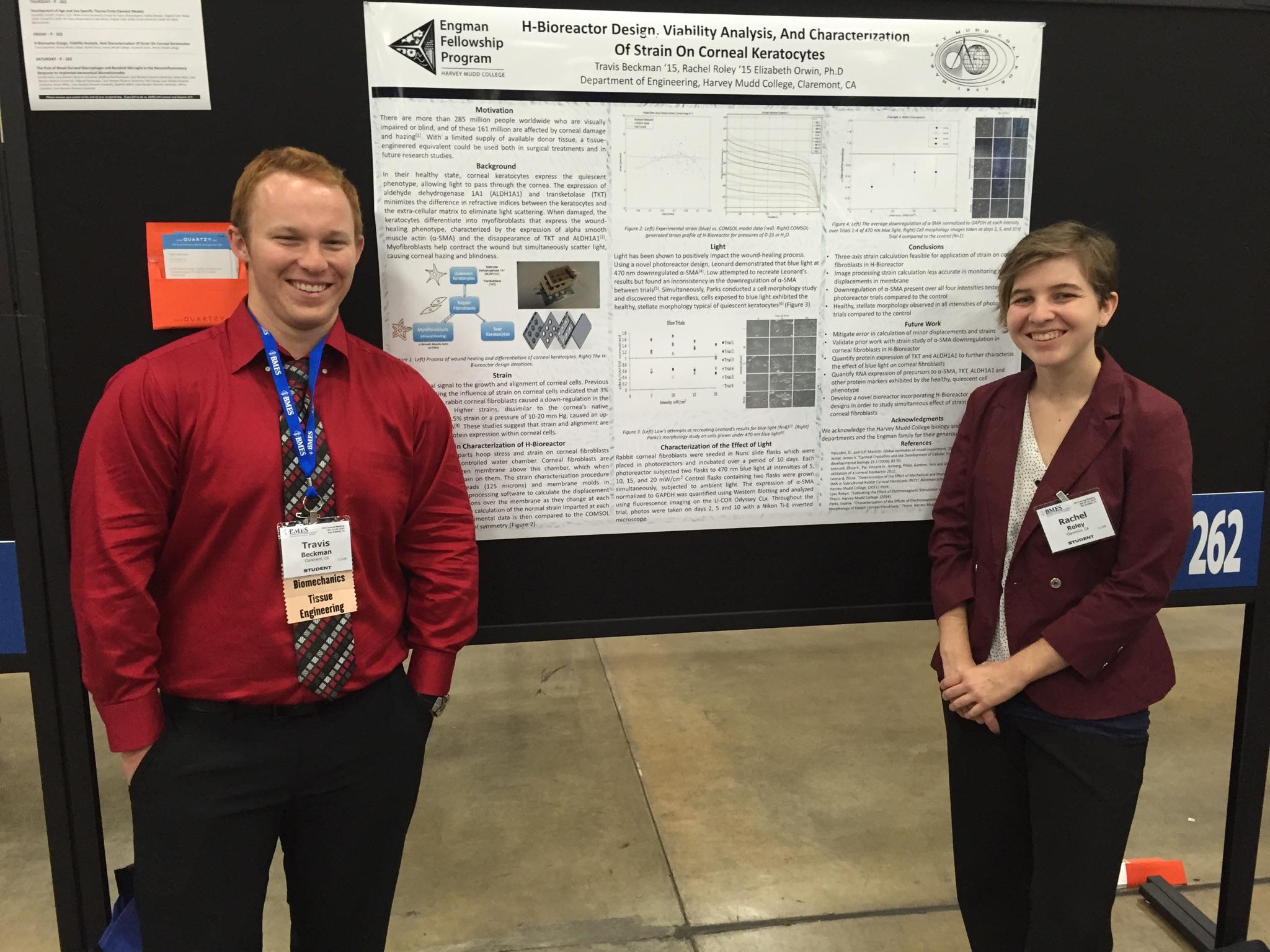
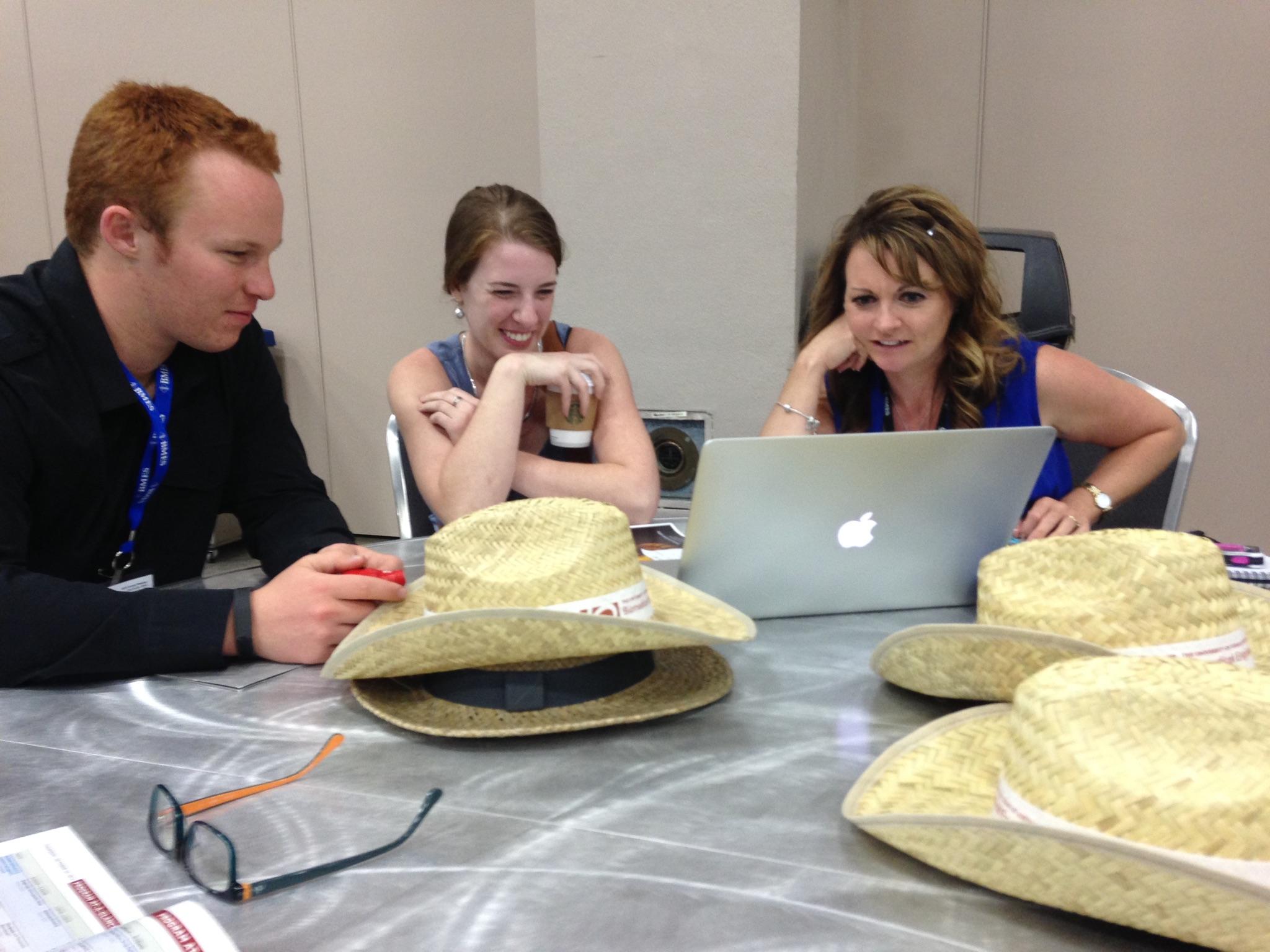
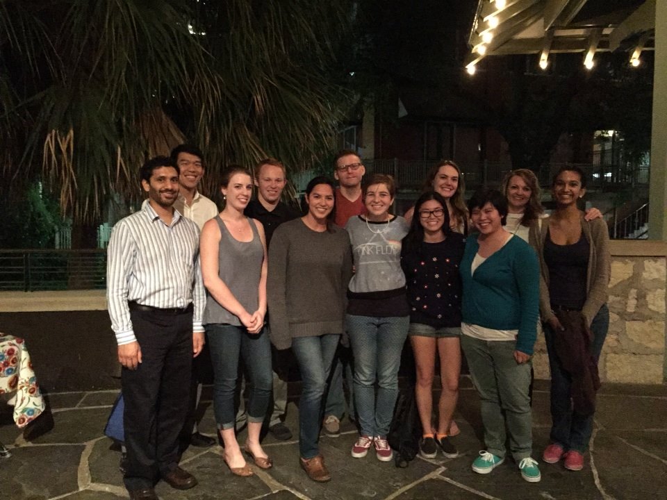
From October 22nd through 25th, seniors Cindy Angpraseuth, Jennifer Zheng, Risa Egerter, Rachel Roley, and local high schooler Kavi Curlin traveled to San Antonio, Texas for the 2014 Biomedical Engineering Society Conference. “It was one of the most formative career experiences for me,” says Travis, “and I’m very thankful that I’m in a school and a lab that allows me to have these kinds of experiences.” The students had the opportunity to network with industry professionals, professors from graduate schools they are applying to, and even Mudd alumni, some of whom are alums of the lab itself. “We’ve built an ‘army’ of people doing cool things in biomedical engineering,” laughs Prof. Orwin, describing her growing group of former lab members. With the interesting techniques of the cornea team and the culturing of astrocytes for gels by the brain patch team, Risa concludes, “Overall, I would say [the research] was well received. The moderator for the poster session even came by and congratulated us on being there!” In the end, the conference proved to be a beneficial experience for the students, the lab, and their research.